The 6 main types of basal cell carcinoma and how they differ
What is basal cell carcinoma?
Basal cell carcinoma (BCC) is the most common, locally invasive, keratinocytic non-melanoma skin cancer. It is also known as rodent ulcer. It rarely spreads to other parts of the body or kills but it can cause significant destruction and disfigurement by invading surrounding tissues. Subtypes of basal cell carcinoma may differ in appearance, aggressiveness, treatment options, and who’s likely to develop them.
What are the types of BCC?
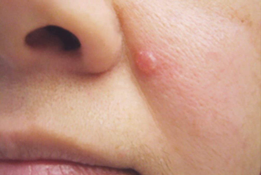
Nodular BCC
- Most common type
- Commonly on the face
- Shiny or pearly lump with a smooth surface
- May have central ulcer
- Usually grows slowly
- Blood vessels cross its surface
- Usually treated surgically
Superficial BCC
- Most common type in younger adults
- Often multiple
- Most common type on upper trunk and shoulders
- Scaly, irregular plaque
- Bleeds or ulcerates
- Can be treated with topical creams such as Effudix or Imiquimod or cryotherapy
Morphoeic BCC – also known as sclerosing or infiltrating basal cell skin cancer
- Usually found in mid-facial sites
- Waxy, skin-coloured, scar-like plaque with indistinct borders
- May infiltrate cutaneous nerves (perineural spread)
- Prone to recurrence after treatment
- Its treatment typically involves surgical procedure such as MOHS
Basosquamous carcinoma
- Mixed BCC and SCC
- Infiltrative growth pattern
- Potentially more aggressive than other forms of BCC
- Also known as basosquamous carcinoma and mixed basal-squamous cell carcinoma
- It’s more likely to spread to other body parts and recur
Micronodular BCC
- Made of tiny nodules that can extend more widely under the surface than expected
- Because edges are harder to judge, it is treated as higher risk for recurrence
- It’s often found on the back as light yellowish lesions that do not ulcerate
- Treatment may include Mohs surgery due to its precision in removal
Pigmented BCC
- Brown, blue or greyish
- May resemble malignant melanoma
- This form of skin cancer can sometimes be more challenging to diagnose as it may resemble other pigmented lesions.
- Treatment options may include surgical excision or Mohs surgery
What are the aggressive forms of basal cell carcinoma?
The aggressive forms of BCC include:
- Infiltrative
- Morpheaform
- Basosquamous
- Micronodular
These types are more likely to invade surrounding tissues and have a higher risk of recurrence than other forms of basal cell carcinoma.
How is BCC diagnosed?
Diagnosis of BCC is based on clinical features. To confirm the diagnosis, a small piece of the abnormal skin (a biopsy), or the whole area (an excision biopsy), is removed under a local anaesthetic and sent to a pathologist to be examined. Some typical superficial BCCs on trunk and limbs are clinically diagnosed and have non-surgical treatment without biopsy.
Based on the results, they can classify the type of BCC and develop an appropriate treatment plan.
How can BCC be treated?
If caught early, BCC is curable and cause minimal damage. However, the larger and deeper a tumor grows, the more dangerous and potentially disfiguring it may become, and the more extensive the treatment must be.
The treatment used will depend on the type, depth of penetration, size and location of the BCC, as well as the patient’s age and general health.
Most BCCs (nodular, infiltrative, morphoeic) are treated surgically. This involves removing the BCC with a margin of normal skin around it (3-4mm), using a local anaesthetic. The skin is then closed with stitches or defect is reconstructed with a local flap or skin graft.
Sometimes other surgical methods are used such as:
Mohs Surgery
It is done by a physician trained in Mohs micrographic surgery. While the patient waits, frozen sections of this excised layer are mapped in detail and examined under a microscope, generally in an on-site laboratory. If cancer is present in any area of the excised tissue, the procedure is repeated only on the body area where those cancer cells were identified (the tissue mapping allows the Mohs surgeon to pinpoint this area of the body), until the last excised layer viewed microscopically is cancer-free. This technique can save the greatest amount of healthy tissue and has the highest cure rate, 99 percent or better. It is used in high-risk areas of the face around eyes, lips and nose.
Curettage and cautery
It is suitable for small, well defined nodular or superficial lesions. This involves scraping the BCC away under local anaesthetic. Wound is left open to heal by itself.
Cryotherapy (freezing)
It is reserved for small, superficial BCCs on trunk and limbs. The tumor tissue is destroyed by freezing it with liquid nitrogen, using a spray device. Later, the lesion and surrounding frozen skin may blister or become crusted and fall off, usually within weeks. The procedure may be repeated several times at the same session to help ensure destruction of all malignant cells. Redness, swelling, blistering and crusting can occur following treatment. Leaves a permanent white mark.
Topical anti-cancer ointments
These ointments include 5-fluorouracil (5-FU) and imiquimod for treatment of superficial BCC. Imiquimod stimulates the immune system to produce interferon, a chemical that attacks cancerous and precancerous cells, while 5-FU is a topical form of chemotherapy that has a direct toxic effect on cancerous cells. Imiquimod is applied 3-5 times each week for 6-16 weeks while f-FU requires prolonged course twice daily for 6-12 weeks. It can take up to 12 weeks to fully settle down.
Photodynamic therapy
It may be used for some superficial BCCs but is best avoided if tumour is in site at high risk of recurrence.
Radiotherapy
It is mainly used if surgery is not suitable. Can’t be used in Gorlin syndrome.
Combined therapy
For advanced BCC, BCC that is recurrent or has spread to other parts of the body a combination of surgery, radiotherapy and targeted therapy is used. That is discussed by a multidisciplinary team of specialists. Oral drugs for advanced or metastatic BCC include Vismodegib and Sonidegib, which are oral hedgehog inhibitor drugs. They have significant risks and side effects.
It is important to note that (unlike Mohs surgery and excisional surgery), curettage and electrodesiccation, radiation, cryosurgery, and topical medications all have one significant drawback in common – since no tissue is examined under the microscope, there is no way to determine how completely the tumour was removed.
Takeaway
BCC types include nodular, superficial, morpheaform, and pigmented, each with different characteristics. Early detection may increase the chance of curing the condition, and treatment options may vary from surgical excision to topical therapies.
Recent Comments